Gallbladder
A pear-shaped reservoir under the liver with a fundus, body, neck and infundibulum. It releases bile when the gut signals after food.
 Request consultation
The gallbladder surgery guide
A cholecystectomy removes the gallbladder—not the liver and not the common bile duct.
The liver continues to make bile, which flows directly into the intestine after the gallbladder is removed.Start with the anatomy
The liver makes bile. The gallbladder stores and concentrates it between meals. The cystic duct connects the gallbladder to the common hepatic duct; below that junction, the channel is called the common bile duct.
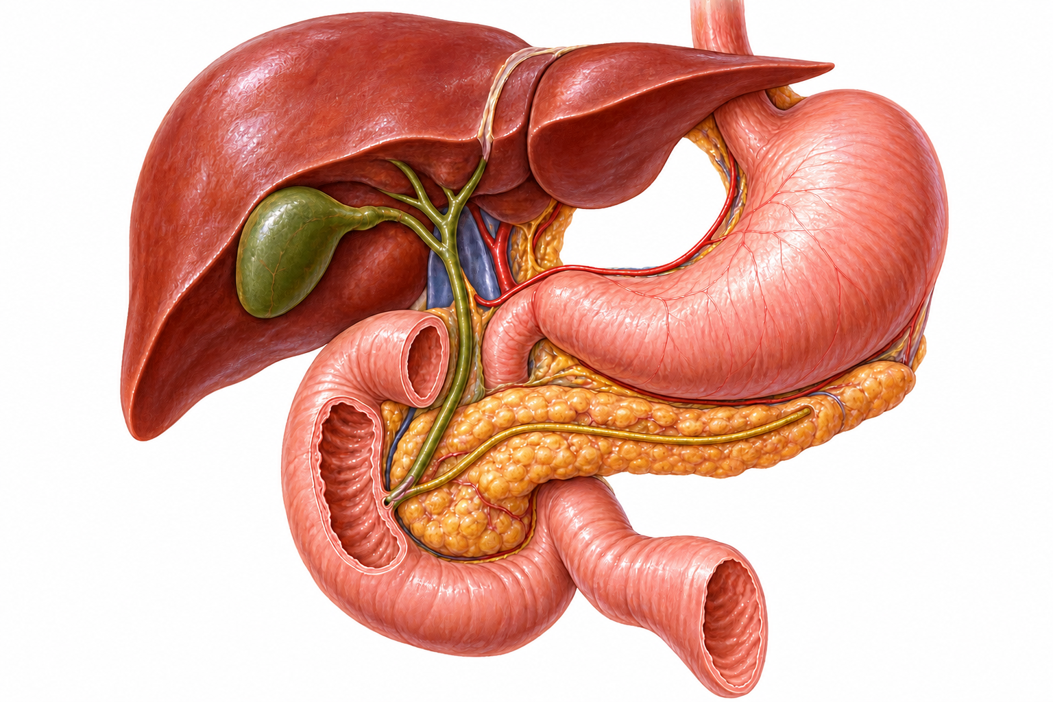
Regional anatomy
An anterior-oblique view establishes how the biliary system sits within the upper abdomen.
Magnified biliary anatomy
The close-up separates the gallbladder, hepatic ducts, CBD and pancreatic outlet from the surrounding regional anatomy.
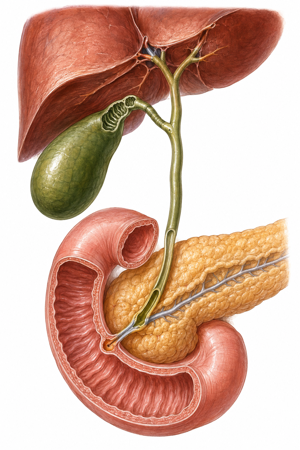
A pear-shaped reservoir under the liver with a fundus, body, neck and infundibulum. It releases bile when the gut signals after food.
The short channel from the gallbladder. It usually joins the common hepatic duct, but length, course and junction vary between people.
The main drainage channel to the duodenum. It is close to the operative area but is not a structure removed or clipped during routine cholecystectomy.
Inflammation can pull, scar or visually distort these structures. Safe surgery requires certainty about anatomy before anything is divided.
Conditions affecting the gallbladder
Symptoms and management depend on where a stone is, whether inflammation or infection is present, and whether the bile duct or pancreas is involved.
Stones without symptoms
Gallstones found incidentally do not automatically require surgery. Observation is common unless symptoms, complications or a specific high-risk context changes the balance.
Temporary blockage
A stone temporarily blocks the cystic duct, causing steady upper-right or upper-middle abdominal pain—often after food—without established gallbladder inflammation.
Sudden inflammation
Persistent cystic-duct obstruction triggers inflammation. Pain usually lasts longer, and fever, tenderness, raised inflammatory markers or supportive scan findings may appear.
Long-standing change
Repeated irritation can leave the gallbladder scarred, thick-walled or contracted. Symptoms may be recurrent or less dramatic than an acute attack, but surgery can be technically demanding.
Pus under pressure
An infected, obstructed gallbladder fills with purulent material. This is a complicated form of acute cholecystitis requiring urgent hospital treatment and source control.
Complicated inflammation
Severe inflammation can compromise the gallbladder wall’s blood supply, causing tissue death, leakage or a local abscess. These are urgent complications—not routine stages every patient passes through.
Choledocholithiasis
A stone leaves the gallbladder and enters the common bile duct. Jaundice, abnormal liver tests, cholangitis or pancreatitis may follow, and the duct may need separate treatment such as ERCP.
Pancreatic inflammation
A migrating stone can transiently block the shared outlet of the bile and pancreatic ducts. Management addresses the pancreatitis, any retained duct stone and the gallbladder.
Wall findings
Many small polyps are benign cholesterol deposits. Adenomyomatosis is usually benign wall thickening. Size, shape, growth and patient risk factors determine follow-up or surgery.
When not to wait
Seek urgent hospital assessment for:
Inflammation & infection
Biliary colic is temporary obstruction without established inflammation. Acute cholecystitis is inflammation; bacterial infection can complicate it. Chronic cholecystitis is a long-standing structural process, not simply “the next stage.”
Tokyo severity grading
Grades I–III describe the severity of an acute episode and help guide timing, expertise and supportive care. They are not cancer stages.
Grade I
Grade II
Grade III
Acute cholecystitis
Typical clues include sustained pain, local tenderness, fever or inflammatory blood-test changes plus supportive imaging. Wall thickening is only one possible sign.
Chronic cholecystitis
The gallbladder may become fibrotic, contracted and densely adherent after repeated irritation. Symptoms can recur without the systemic illness of an acute episode.
Empyema
Pus accumulates inside an obstructed gallbladder. Patients can deteriorate quickly and need urgent antibiotics, drainage or surgery according to fitness and severity.
Understanding the ultrasound report
A scan finding is evidence—not a diagnosis by itself. Symptoms, examination, blood tests, gallbladder distension, stones, fluid and bile-duct findings must be interpreted together.
Gallbladder wall
A properly distended gallbladder wall is generally considered thickened above 3 mm. Acute or chronic cholecystitis can cause thickening, but so can hepatitis, heart or kidney failure, low albumin, ascites and other systemic conditions.
Gallbladder polyps
Most small polyps are benign. Using the 2022 European joint guideline as an orientation, management becomes more active when a lesion is 10 mm or larger, or when a 6–9 mm lesion accompanies risk factors.
Risk factors used with a 6–9 mm polyp
These thresholds guide a clinical conversation; societies differ, ultrasound can overcall pseudopolyps, and suspicious mass-like or invasive features need an HPB/oncology pathway rather than routine polyp surveillance.
Cancer risk in perspective
Gallstones and chronic inflammation are associated with gallbladder cancer, but gallstones are common and gallbladder cancer is rare. Larger or changing polyps, a sessile lesion, irregular focal thickening, a mass, liver invasion or suspicious nodes deserve more concern. Removed gallbladders are examined by pathology; an unexpected cancer finding may require staging and further HPB surgery.
Management options
The correct pathway depends on symptoms, inflammation, bile-duct involvement, pancreatitis, polyp features, cancer suspicion and the patient’s fitness for an operation.
Often appropriate for symptom-free gallstones and some low-risk small polyps. New symptoms or interval scan changes should trigger reassessment.
Commonly considered for recurrent biliary colic, chronic cholecystitis, previous complications and selected gallbladder polyps.
Hospital care may include fluids, pain control, antibiotics when indicated and early laparoscopic cholecystectomy for suitable patients.
MRCP or EUS can clarify suspected common bile duct stones. ERCP removes duct stones but does not remove the gallbladder itself.
Percutaneous or endoscopic drainage may control infection in selected critically ill or very high-risk patients when immediate surgery is unsuitable.
A suspicious mass, complex duct problem, bile duct injury or unexpected cancer requires dedicated staging and specialist hepatopancreatobiliary planning.
A useful distinction
ERCP treats the main bile duct. A cholecystostomy drains the gallbladder. A cholecystectomy removes the gallbladder. They solve different parts of biliary disease and may sometimes be used in sequence.
Gallbladder removal
The operation disconnects the gallbladder from its duct and blood supply, separates it from the liver bed and removes it—usually through small laparoscopic ports.
A camera and long instruments are introduced through several small ports.
The standard minimally invasive route for most routine and many acute cases.
Small incisions do not remove the need for meticulous duct identification.
The surgeon controls wristed instruments and a magnified 3D camera from a console.
Selected anatomy or complex minimally invasive dissection where the platform adds value.
The robot does not operate autonomously and is not universally superior.
The gallbladder is removed through an incision beneath the right rib margin.
Selected severe inflammation, complex anatomy, previous surgery, cancer concern or conversion for safety.
Conversion is a judgement made to protect the patient—not a complication by itself.
Part of the gallbladder is deliberately left when complete dissection is unsafe.
A bailout option when the cystic duct and artery cannot be safely defined.
It can reduce major duct-injury risk but has its own follow-up considerations, including bile leak or retained stones.
The operation, simplified
After the Critical View of Safety is established, the cystic structures are sealed and divided. The gallbladder is then freed from the liver and removed in a retrieval bag.
Small clips commonly remain on the patient side of the cystic duct and artery. A clip on the gallbladder side leaves with the specimen. Clip number and alternatives vary; the principle is secure closure after correct identification.
The consultation
A gallbladder consultation should connect the symptoms, blood tests and imaging; assess the bile duct and pancreas when relevant; explain whether observation, ERCP, drainage or surgery is appropriate; and discuss the planned approach plus safety alternatives.
Request a gallbladder consultationCommon patient questions
The gallbladder is the site where stones form. Removing stones alone leaves the diseased gallbladder behind, so stones and symptoms can recur. Cholecystectomy treats the source while the liver continues to produce bile.
Yes. The liver keeps making bile, which flows directly through the hepatic ducts and common bile duct into the small intestine. Most people return to a normal diet and daily life after recovery.
No. Gallstones that have never caused symptoms are often observed. Surgery is considered differently when there is biliary pain, inflammation, pancreatitis, jaundice, a duct stone or another complication.
Blood tests and ultrasound may raise suspicion; MRCP or EUS can define the duct. A confirmed stone may be removed by ERCP or, in selected settings, bile-duct exploration during surgery. Cholecystectomy and duct clearance are related but separate decisions.
Small surgical clips commonly remain on the patient side of the cystic duct and cystic artery. Alternatives include sutures, energy-sealing devices or stapling in selected circumstances. Your surgeon chooses the safest method for the anatomy encountered.
No. Laparoscopic cholecystectomy remains the standard minimally invasive operation. Robotic assistance may be useful in selected cases, but it is an access platform—not a guarantee of a safer or better result.
Safe surgery means not dividing uncertain structures. The surgeon may pause, use imaging, change the direction of dissection, perform a subtotal cholecystectomy, convert to open surgery or seek specialist help. A planned bailout is a safety decision, not a failure.
Usually not. A lighter diet can be comfortable early in recovery, then foods are reintroduced according to tolerance. A small number of people notice temporary softer or more frequent stools.
Many uncomplicated laparoscopic cases go home the same day or after a short stay and resume light activity early. Work, driving, lifting and exercise timelines depend on the operation, inflammation, complications and the individual patient.
No. Gallstones are common and gallbladder cancer is rare; most small polyps are non-cancerous. Concern rises with features such as a larger or sessile polyp, focal irregular wall thickening, growth, a mass, primary sclerosing cholangitis or older age. Suspicious findings need specialist review rather than assumptions.
Medical references
Patient information informed by the NIDDK gallstone treatment guide and American College of Surgeons cholecystectomy guide. Anatomical relationships reference the NCBI gallbladder anatomy review and NCBI biliary-duct review. Surgical-safety content follows the SAGES multi-society Safe Cholecystectomy guideline and SAGES biliary surgery guidance. Acute cholecystitis content references the Tokyo Guidelines 2018 and 2020 WSES guideline. Polyp management references the 2022 ESGAR/EAES/EFISDS/ESGE joint guideline.
Reviewed for website publication in August 2026. This guide is educational, does not diagnose gallbladder disease and cannot replace individual surgical assessment.